Call Us - (+20) 122 808 5310

ultrasound examination
Ultrasound 3D and 4D
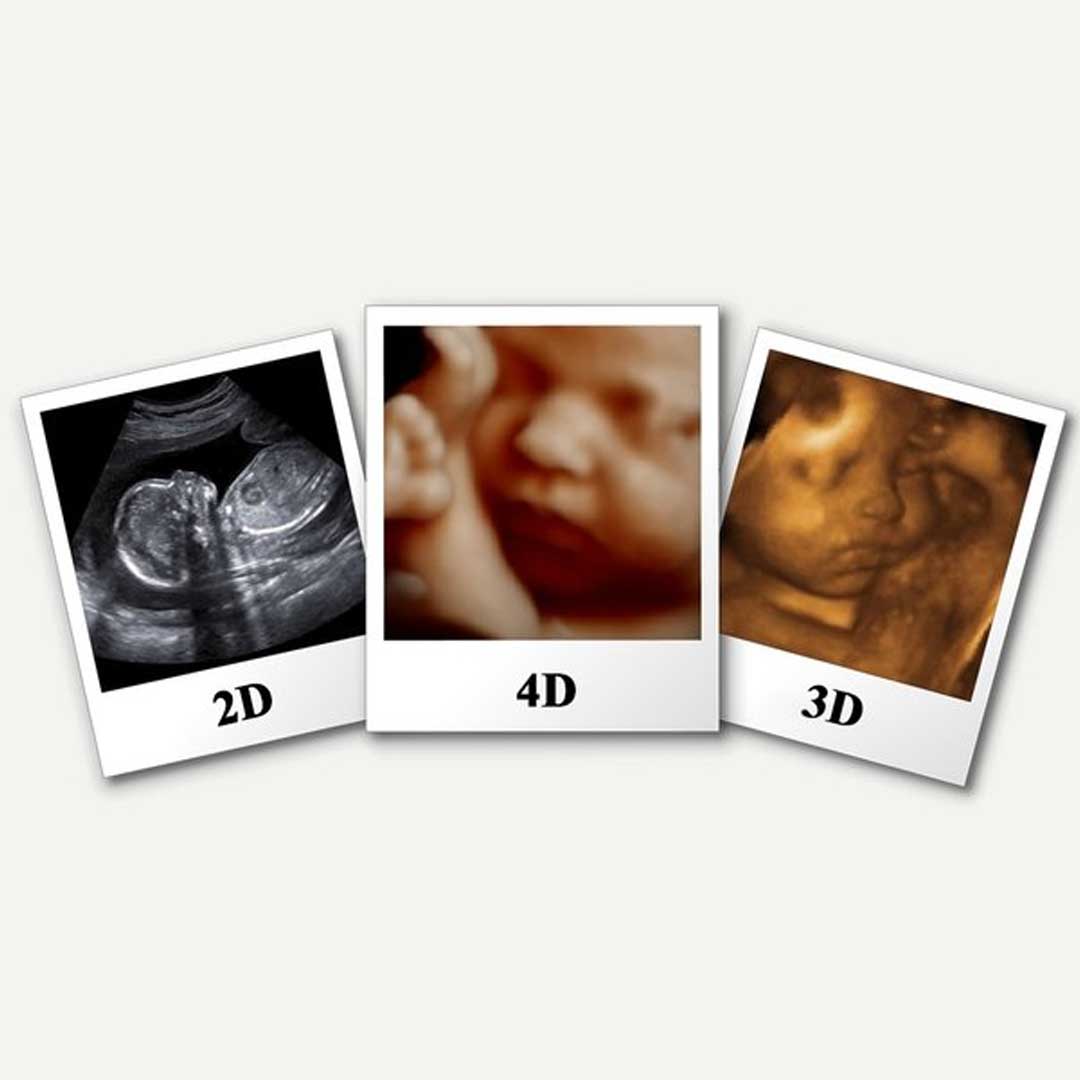
Advanced imaging methods like as ultrasound 3D and 4D enable real-time, three-dimensional pictures of the fetal anatomy and movements.
These techniques can yield important data for evaluating the health of the fetus and identifying any potential defects or pregnancy-related issues. It is advised to use ultrasound 3D and 4D as supplementary instruments to the conventional 2D ultrasound.
Make an Appointment3D Ultrasound Examination
finds any possible issues in the first several months of pregnancy.


Ultrasound 4D Examination
makes a video clip of the fetus's motions to better display the baby's face, features, and movements. The technology used is also different. The majority of the time, 4D ultrasound is utilized between weeks 20 and 24, although occasionally, if a doctor suspects that the fetus has defects, they may request one as early as week 11.
Frequently Asked Questions
How significant is a 4D ultrasound?
For certain women, a 4D ultrasound can be an invaluable tool. Parents may find it comforting to have a closer look at the baby’s anatomy and movements. Additionally, it can be used to identify certain birth abnormalities.
A 4D ultrasound: Is it Safe?
The doctor in charge of the prenatal care should be the one to decide whether or not to utilize 4D ultrasound since only they are qualified to determine whether this method is necessary given the fetal status.
In what ways do 4D and 3D ultrasounds vary?
While 4D ultrasounds can display your baby’s activities, 3D ultrasounds can only show you how your baby looks.
Your requirements and tastes will determine which kind of ultrasound is ideal for you. Three-dimensional ultrasound could be a fantastic choice for you if you want to get an intricate picture of your unborn child’s anatomy. An improved choice would be a 4D ultrasound if you want to view your baby move.

